Our research portfolio keeps expanding. This is the best time for medical physics research and discovery. Stay tuned for the announcement of exciting new research directions!
4π Radiotherapy
4π Radiotherapy
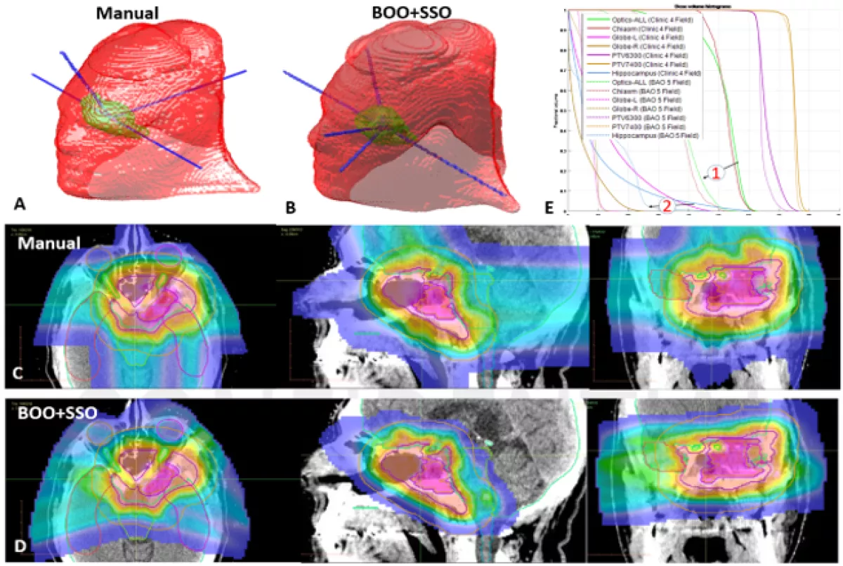
With the exception of intracranial treatment, radiotherapy has been largely coplanar, which is easier to deliver but results in suboptimal dose distribution. We develop optimization, 3D modeling and navigation schemes to solve the challenging problems and show the feasibility of creating and deliver highly non-coplanar 4π plans on a new linac.
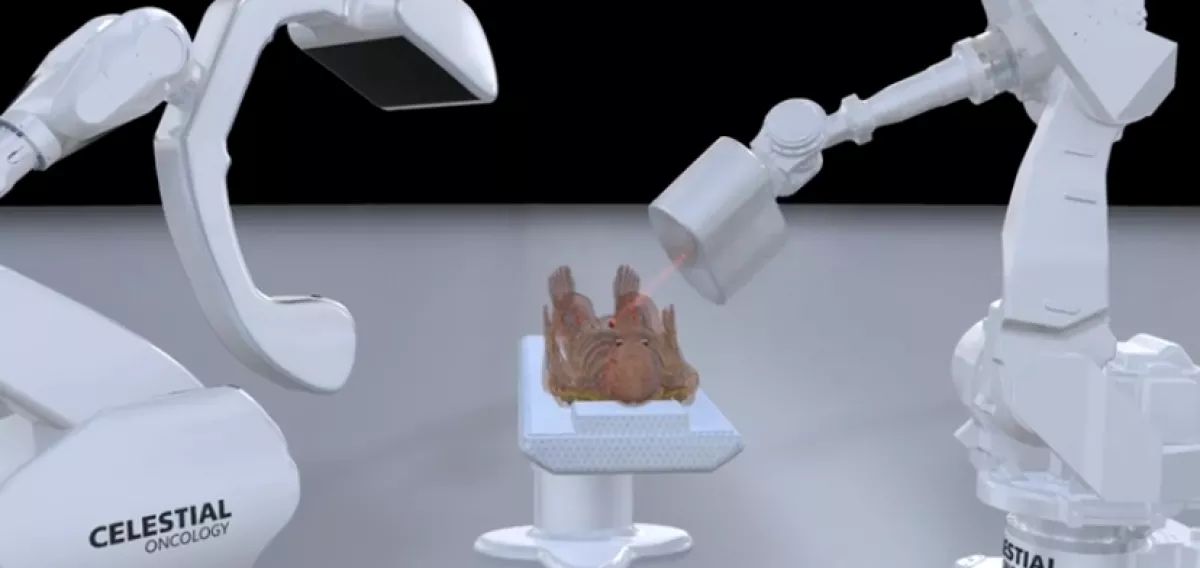
Robotic radiotherapy system under construction at UCLA and Celestial Oncology
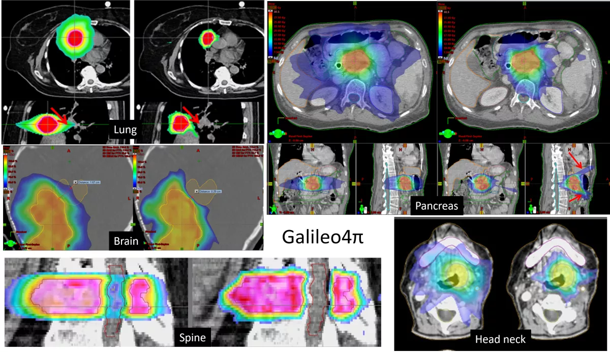
Dosimetric examples of using highly non-coplanar beams to markedly reduce normal tissue dose
MRI Guided Radiotherapy
MRI Guided Radiotherapy
MRI is playing an increasingly important role in radiotherapy by providing kenetic, functional and real time monitoring of the tumor and normal tissue for improved radiotherapy accuracy and efficacy. We are interested in solvng problems assocaited with MRI guided radiotherapy, including fast MRI acquisition, automated organ delineation, deformable registration and utilization of functional MRI information for adaptive radiotherapy and outcome assessment.

Reconstruction of the coronal lung MRI scan using different algorithms: frame 1, 10 and 19 and the temporal profile (left to right). Top row: fully sampled MRI sequence with ROI contoured using red dashed rectangle and the location of the extracted temporal profile indicated using blue vertical line. Bottom rows: zoomed spatial ROI of the reconstructed MRI scans using ktSLR, L+S, MaSTER and the proposed MC-JPDAL.

Ultimate form of MRgRT with real time imaging, adaptive planning and delivery
Machine learning and AI
Machine learning and AI for automated segmentation, registration, Monte Carlo dose calculation, dose and treatment plan prediction
Machine learning and AI have seen a rapid growth in recent radiotherapy research. We are particularly interested in using these methods for image segmentation, registration, dose calculation, dose prediction and automated plan generation.

Examples of the segmentation results by ResNet, DeepLab V3+, FCNN-BSR, HMEDN, and the proposed MTER-Net. The first row shows the ground truths, the second to sixth rows present the segmentation results by ResNet, DeepLab V3+, FCNN-BSR, HMEDN, and the proposed MTER-Net, respectively. Bladder-red, Rectum-green, Prostate-yellow, Seminal Vesicle-blue. The dashed circles indicate the regions with significant improvements.
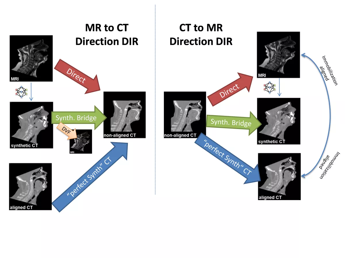
The deformable registrations performed in this study. Registrations were performed in both CT-to-MR and MR-to-CT directions to test for inverse consistency, as well as both directly (multi-modal) and with a synthetic CT bridge (synthetic mono-modal). The DVF from CTsynth CTnon-aligned was applied to the MR to generate a deformed MR image. The non-aligned CT was also registered to the aligned CT (and vice versa) to see an approximate best-case synthetic mono-modal registration.
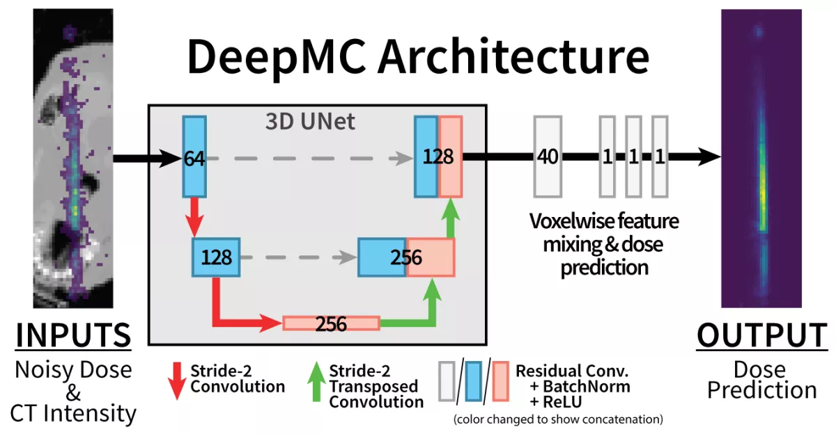
Fully convolutional model architecture used by our method, DeepMC. Numbers in blocks indicate number of feature channels produced by learned convolutional kernels at each stage. 3×3×3 kernels are used in UNet layers. 1×1×1 kernels are used for feature mixing and dose prediction.
Development of the next generation FLASH X-ray system
Development of the next generation FLASH X-ray system
Ultra-high dose rate radiotherapy, a.k.a. FLASH radiotherapy, has shown the potential to drastically reduce the normal tissue toxicity without compromising tumor cell killing. However, the delivery of FLASH is hampered by the limitations inherent to existing radiotherapy platforms. We are developing a groundbreaking new X-ray system, ROAD, to achieve the needed FLASH dose rate for significantly improved cancer radiotherapy efficacy.

Four beams selected from a ROAD delivery sequence with respective MLC apertures in the upper left corner and cumulative isodose distribution for a brain patient. In this configuration, the MLC-ring fits 75 MLC modules with 15cm beam width in the isocenter plane. With static MLC-ring, by rotating the source for one round, the source is sequentially aligned with all 75 MLC openings, achieving 75 beams within one rotation (ROAD-75). By rotating the source and the ring-collimator in the opposite direction with the same speed, the source-MLC aligning positions are doubled, achieving 150 beams with one round of source rotation and collimator rotation (ROAD-150).
Proton and heavy charged particle treatment planning optimization
Proton and heavy ion treatment planning optimization
Proton and heavy charged particles have distinctly different physics compared with X-rays for radiotherapy. Most notably, they form Bragg peaks that are particularly desirable for the radiation dose distribution. Meanwhile, there are remaining challenges in proton and heavy ion treatment planning delivery. We aim to solve the beam orientation optimization, robustness and RBE problems associated with these modalities.
Automated beam orientation based on non-convex term regularized optimization result in less operator-dependent superior proton dosimetry.

SenR is a new proton robustness regularization method that comprises less the dosimetry yet is more resilient to larger range uncertainties.

By incorporating LET into treatment planning, normal tissues are better protected from beams that are potentially highly radiobiologically effective (more toxic).
CT Reconstruction
Dual and single energy CT reconstruction
Medical image acquisition and reconstruction are an integral part of radiotherapy. We are particularly interested in solving the image reconstruction problem as a convex and non-convex optimization problem. For example, we formulate the dual energy CT multiple material decomposition problem as a non-convex optimization problem. We simultaneously solve multiple materials while at the same time suppressing the noise that commonly and adversely affects the utility of dual energy CT. The versatile regularization can be extended to low dose CT reconstruction and other applications.
Decomposition component images of (1) bone (2) iodine (3) muscle (4) fat and (5) air, decomposed using the proposed framework when (a) α ¼ 0, (b) α ¼ 12, (c) α ¼ 23, and (d) α ¼ 1; (e) the DI method. The last column is the 5 × 5 NCC map of the decomposition in the same row, with each square showing the corresponding entries in the NCC matrix, where the basis materials are bone, iodine, muscle, fat, and air, from top to bottom and from left to right.

A classical optimization method ADMM can be reformulated to incorporate flexible image denoiser such as BM3D and even deep learning neural networks for superior low dose CT reconstruction performance.
Small animal dosimetry
Small animal dosimetry
Preclincial radiation is indispensable to test hypotheses, methods, and interventions before human trials. However, there are large disparities in the dosimetric quality and accuracy between human therapy and animal studies. We endeavored to fill the gap and make preclinical experiments more translatable to human patients.

Comlex simultaneous integrated boost plan for mice based on PET uptake using the Sparse Orthogonal Collimator.

3D printed non-human primate phantom with ceramic bones, low and high density soft tissues

Each TLD is different. By tracking them based on their unique appearance, robust individual TLD registration and dosemetry can be achieved via fingerprinting.
Pair Production Tomography
Pair Production Tomography
MV X-rays interaction with tissues and matters can also produce positrons that would annihilate, resulting in a pair of photons, which if detected, can be used to reconstruct tomographic images that are uniquely useful for diagnosis and image guided radiotherapy.
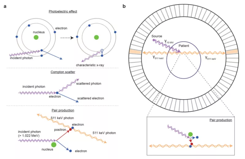
Figure showing how pair production tomography (P2T) images are formed
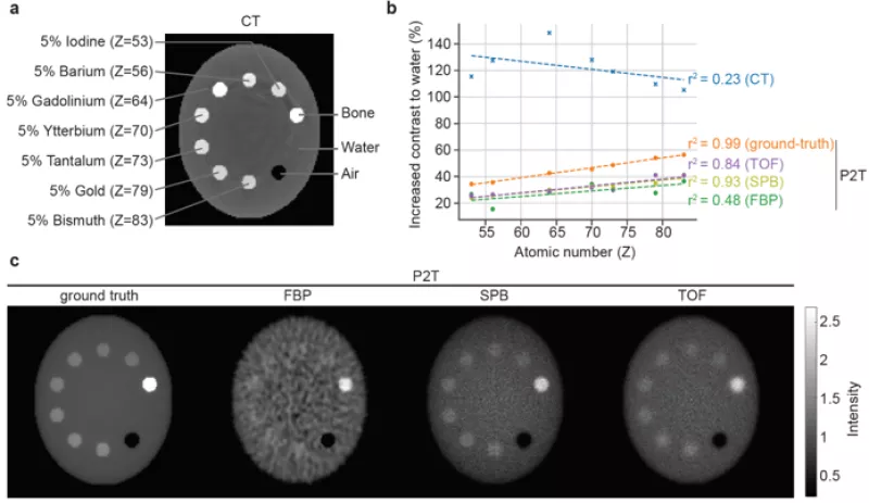
Compared with CT, P2T contrast is linear to the material atomic number, which an desirable property for material differentiation
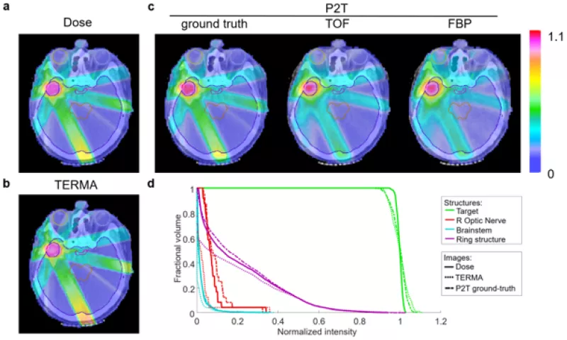
The P2T signals directly reflect the interaction between MV X-rays with the tissue and is an excellent surrogate for in vivo dosimetry
Brachytherapy
- Analytical HDR Prostate Brachytherapy Planning with Automatic Catheter and Isotope Selection, C Holly Frank, Pavitra Ramesh, Qihui Lyu, Dan Ruan, Sangjune Park, Albert Chang, Puja Venkat, Amar U Kishan, Ke Sheng, Medical Physics 2023 http://doi.org/10.1002/mp.16677
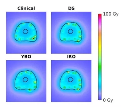
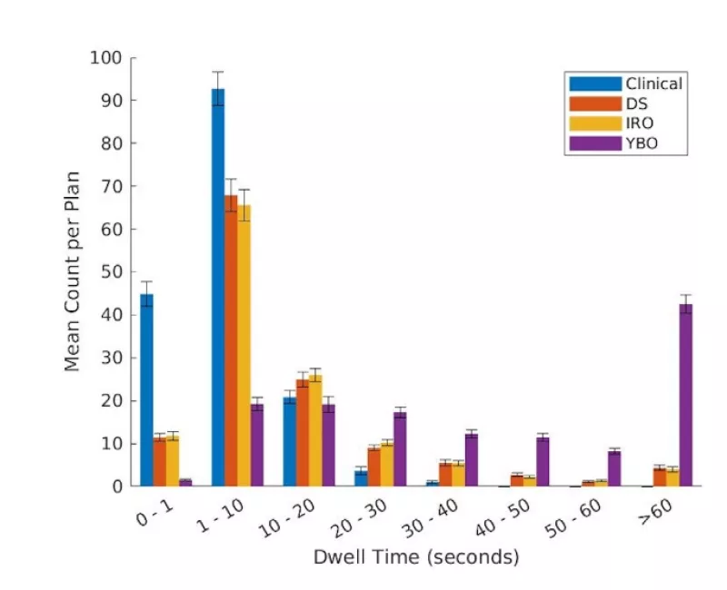
Axial dose distribution for an example patient. Population averaged dwell time histogram withstandard error bars for the clinical, DS, IRO, and YBO plans.
Ultra-large digital tumor model
- Gell: A GPU-powered 3D hybrid simulator for large-scale multicellular system, Jiayi Du, Yu Zhou, Lihua Jin, Ke Sheng, Plos ONE, 2023 Jul 18;18(7):e0288721. doi: 10.1371/journal.pone.0288721